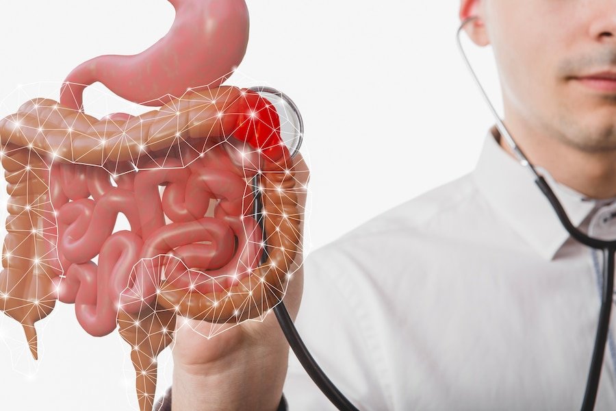
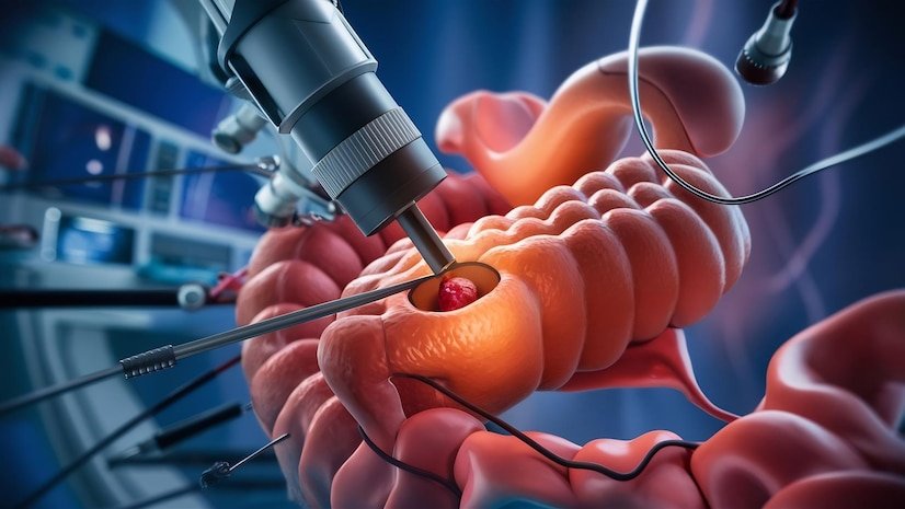
Are you in search of a reliable and experienced Colonoscopy Treatment Specialist in Prashant Vihar? Look no further, as Dr. Alok Tiwari at dralokitgulatigastro.com is your go-to expert for all your colon health needs—Colonoscopy Treatment Specialist in Prashant Vihar. To Know More About It Please Click Here Colonoscopies are vital procedures that aid in the diagnosis and treatment of various gastrointestinal conditions. Whether you’re experiencing symptoms like abdominal pain, rectal bleeding, or changes in bowel habits, or you’re due for routine screening, having a skilled specialist by your side is essential. Dr. Alok Tiwari stands out as a leading Colonoscopy Treatment Specialist in Prashant Vihar, with years of expertise and a commitment to providing personalized care to each patient. Let’s delve into why he’s the top choice for your colon health needs. Expertise: Dr. Alok Tiwari boasts extensive experience in performing colonoscopies and treating a wide range of gastrointestinal disorders. His expertise ensures accurate diagnosis and effective treatment strategies tailored to your specific condition. Advanced Technology: At dralokitgulatigastro.com, you’ll find state-of-the-art facilities equipped with advanced technology for precise and minimally invasive colonoscopy procedures. Dr. Alok Tiwari utilizes the latest tools and techniques to ensure optimal outcomes for his patients. Comprehensive Care: As your dedicated Colonoscopy Treatment Specialist in Prashant Vihar, Dr. Alok Tiwari provides comprehensive care from initial consultation to post-procedure follow-up. He takes the time to understand your concerns, answer your questions, and develop a personalized treatment plan that meets your needs. Patient-Centric Approach: Dr. Alok Tiwari believes in a patient-centric approach to healthcare, where your comfort, safety, and well-being are top priorities. You can trust him to provide compassionate care and support throughout your colonoscopy journey. Preventive Screening: Routine colonoscopies are crucial for early detection and prevention of colorectal cancer and other gastrointestinal conditions. Dr. Alok Tiwari emphasizes the importance of preventive screening and encourages patients to undergo regular colonoscopies for optimal colon health. Efficient Process: At dralokitgulatigastro.com, you’ll experience a streamlined and efficient process for scheduling and undergoing your colonoscopy procedure. Dr. Alok Tiwari and his team ensure minimal wait times and smooth transitions to make your experience as convenient as possible. Continued Support: Beyond the procedure itself, Dr. Alok Tiwari offers continued support and guidance to help you maintain a healthy colon. Whether you have questions about post-procedure care, dietary recommendations, or follow-up appointments, he’s always available to assist you. Positive Patient Experiences: Patient satisfaction is paramount at dralokitgulatigastro.com, where countless individuals have experienced successful colonoscopy procedures under Dr. Alok Tiwari’s care. Their testimonials speak volumes about the quality of service and the level of expertise provided by the team. Community Engagement: Dr. Alok Tiwari is actively involved in raising awareness about colon health and the importance of regular screenings within the Prashant Vihar community. Through educational initiatives and outreach programs, he strives to empower individuals to take control of their gastrointestinal health. To Know More About It Please Click Here Accessible Location: Conveniently located in Prashant Vihar, dralokitgulatigastro.com offers easy access to top-notch colonoscopy services. Whether you’re a local resident or visiting from nearby areas, you’ll find the clinic’s location convenient for all your healthcare needs.Colonoscopy Treatment Specialist in Prashant Vihar.
Best Hepatology Consultant in Rohini.
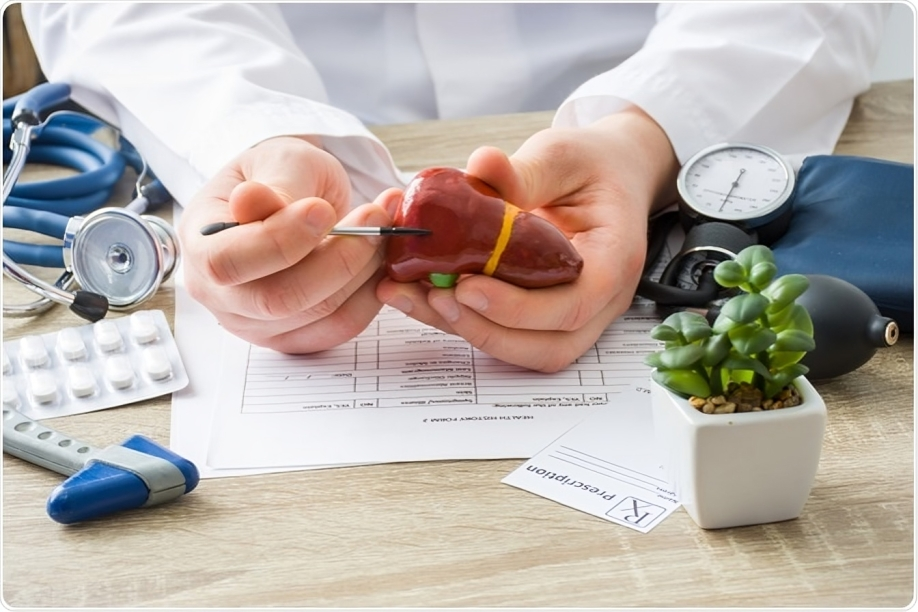
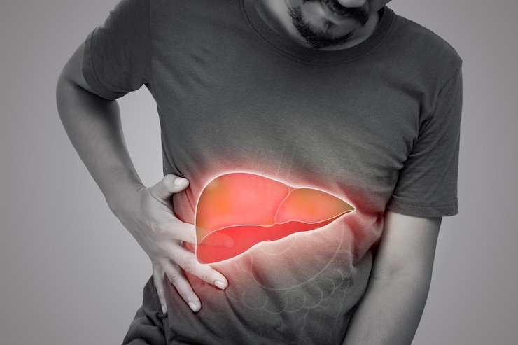
Rohini, known for its vibrant culture and bustling lifestyle, is also home to some of the most esteemed medical professionals in the country. Among its healthcare gems shines a prominent figure in the realm of liver health – the Hepatology Consultant. To Know More About It Please Click Here Exploring the Liver Landscape The liver, a vital organ, performs an array of crucial functions, from metabolizing nutrients to detoxifying harmful substances. However, liver diseases pose a significant threat to public health globally. Amidst this challenge, finding the right Hepatology Consultant becomes paramount. Meet Dr. Aarav Khanna In the heart of Rohini stands Dr. Aarav Khanna, a beacon of hope for those grappling with liver-related ailments. Renowned for his expertise and compassionate care, Dr. Khanna stands as a pillar in the field of hepatology. Education and Expertise Dr. Khanna’s journey towards becoming Rohini’s go-to Hepatology Consultant is adorned with academic excellence and extensive clinical experience. Armed with a stellar medical education and specialized training in hepatology, he brings a wealth of knowledge to the table. Comprehensive Care Approach What sets Dr. Khanna apart is his holistic approach to liver health. Beyond diagnosis and treatment, he emphasizes preventive strategies and patient education. Every consultation with Dr. Khanna is not just a medical encounter but an opportunity for patients to understand their condition and actively participate in their wellness journey. Cutting-Edge Treatments In an ever-evolving field like hepatology, staying abreast of the latest advancements is imperative. Dr. Khanna ensures that his practice integrates cutting-edge treatments and technologies to deliver optimal outcomes for his patients. From advanced diagnostics to innovative therapeutic interventions, his arsenal is equipped to tackle even the most complex liver disorders. Patient-Centric Care At the core of Dr. Khanna’s practice lies a commitment to patient-centric care. He understands that navigating liver diseases can be daunting, often accompanied by emotional distress. Thus, he fosters a supportive environment where patients feel heard, valued, and empowered throughout their treatment journey. To Know More About It Please Click Here Community Engagement and Awareness Beyond his clinical duties, Dr. Khanna actively engages with the community to raise awareness about liver health. Through seminars, workshops, and social media outreach, he endeavors to demystify liver diseases and promote early detection and intervention. ConclusionIn the labyrinth of liver health, Dr. Aarav Khanna emerges as a guiding light for the residents of Rohini. With his blend of expertise, empathy, and innovation, he not only heals bodies but also instills hope and resilience in the hearts of his patients. As Rohini’s finest Hepatology Consultant, Dr. Khanna continues to redefine the standards of liver care, one life at a time.
Best Colonoscopy Treatment Specialist in Rohini.
A colonoscopy is a crucial diagnostic and therapeutic procedure used to detect and treat various gastrointestinal conditions, including colorectal cancer, polyps, inflammatory bowel disease, and gastrointestinal bleeding. If you’re in Rohini and in need of colonoscopy treatment, finding the best specialist is paramount for accurate diagnosis, effective treatment, and personalized care. Here’s a comprehensive guide to help you find the best colonoscopy treatment specialist in Rohini. To Know More About It Please Click Here Research Specialists Begin your search for a colonoscopy treatment specialist in Rohini by researching healthcare professionals who specialize in gastroenterology or gastrointestinal endoscopy. Utilize online search engines, medical directories, and hospital websites to compile a list of potential specialists. Check Credentials and Qualifications Verify the credentials, qualifications, and board certifications of the specialists on your list. Look for specialists who are trained in gastroenterology, have extensive experience in performing colonoscopies, and are affiliated with reputable medical institutions or hospitals in Rohini. Assess Experience and Expertise Evaluate the experience and expertise of each specialist in performing colonoscopy procedures and managing gastrointestinal conditions. Inquire about the number of colonoscopies they perform annually, their success rates, and any specialized training or areas of expertise, such as advanced endoscopic techniques or colorectal cancer screening. Review Patient Reviews and Testimonials Read patient reviews and testimonials to gain insights into the experiences of previous patients with the specialist. Look for positive feedback regarding the specialist’s communication skills, bedside manner, technical proficiency, and overall satisfaction with the colonoscopy procedure and treatment outcomes. Evaluate Facilities and Technology Consider the facilities and technology available at the specialist’s practice or affiliated medical center in Rohini. Look for clinics or hospitals equipped with state-of-the-art endoscopy suites, advanced imaging technology, and facilities for sedation and anesthesia to ensure optimal comfort and safety during the colonoscopy procedure. Inquire About Colonoscopy Preparation Inquire about the colonoscopy preparation process recommended by the specialist and their team. A thorough and personalized preparation regimen is essential for achieving clear visualization of the colon and accurate diagnosis during the procedure. Ensure that the specialist provides detailed instructions and support throughout the preparation process. Ask About Sedation Options Discuss sedation options with the specialist and inquire about their approach to sedation during the colonoscopy procedure. Some patients may prefer conscious sedation or anesthesia-assisted sedation to minimize discomfort and anxiety during the procedure. Choose a specialist who offers sedation options tailored to your preferences and medical needs. Consider Accessibility and Convenience Consider the location, accessibility, and convenience of the specialist’s practice or medical center in Rohini. Choose a location that is easily accessible by public transportation or car, with ample parking facilities and convenient appointment scheduling options to accommodate your needs. Seek Referrals and Recommendations Seek referrals and recommendations from your primary care physician, family members, friends, or colleagues who have undergone colonoscopy treatment in Rohini. Personal referrals can provide valuable insights and help you make an informed decision when selecting a specialist. To Know More About It Please Click Here Schedule a Consultation Finally, schedule a consultation with the selected specialist to discuss your medical history, symptoms, concerns, and treatment options. Use the consultation as an opportunity to ask questions, address any apprehensions or uncertainties, and assess the specialist’s communication style, professionalism, and willingness to listen to your needs. By following these guidelines and conducting thorough research, you can find the best colonoscopy treatment specialist in Rohini who offers personalized care, advanced technology, and expertise in managing gastrointestinal conditions. With the right specialist by your side, you can undergo colonoscopy treatment with confidence, knowing that your health and well-being are in capable hands.
How to Prepare for an Endoscopic Ultrasound Procedure.
An endoscopic ultrasound (EUS) is a minimally invasive procedure that uses sound waves to examine your digestive tract. While it’s a valuable diagnostic tool, some preparation is required to ensure a smooth and successful procedure. Here’s a breakdown of what you can expect. To Know More About It Please Click Here Pre-Procedure Consultation Doctor Discussion: Your doctor will explain the EUS procedure in detail, including its purpose, potential risks, and recovery process. Discuss any questions or concerns you may have.Medical History: Be prepared to share your complete medical history, including any allergies, medications you take (including prescriptions, over-the-counter drugs, and herbal supplements), and past surgeries.Fasting Instructions: You’ll likely be instructed to fast for at least 6-8 hours before the EUS. This ensures your stomach is empty for optimal image quality. Dietary Adjustments Clear Liquids: Depending on the specific location being examined during the EUS, you might need to follow a clear liquid diet for a day or two before the procedure. Clear liquids include water, broth, clear juices (without pulp), and black coffee or tea (without milk or cream).Colon Cleansing: If the EUS will examine your rectum, your doctor may recommend a colon cleansing solution or a combination of a liquid diet and laxatives to clear your bowels. Medication Adjustments Blood Thinners: Inform your doctor about any blood-thinning medications you take, as they may need to be adjusted or stopped temporarily before the EUS to minimize bleeding risks.Continue Certain Meds: In some cases, your doctor may advise you to continue taking specific medications like insulin or certain heart medications with a sip of water even during the fasting period. Day of the Procedure Transportation: Arrange for a ride home after the EUS, as sedation or anesthesia used during the procedure will impair your ability to drive.Clothing: Wear loose-fitting, comfortable clothing that allows easy access to the examination area (usually upper abdomen or rectum).Medications: Take only medications your doctor has specifically instructed you to take on the day of the procedure, with a small sip of water.Additional Tips To Know More About It Please Click Here Inform the Doctor of the Issues If you experience any illness, fever, or other health concerns before your EUS, contact your doctor immediately. They may need to reschedule the procedure.Valuables: Leave valuables at home as you may not be able to keep them with you during the procedure.Questions Welcome: Don’t hesitate to ask your doctor or nurse any questions you may have before, during, or after the procedure.By following these preparation steps and openly communicating with your doctor, you can ensure a smooth and successful endoscopic ultrasound experience.
7 Common Mistakes to Avoid in Spyglass Cholangioscopy.
Spyglass cholangioscopy is a valuable diagnostic and therapeutic tool used in the management of biliary disorders, offering detailed visualization of the bile ducts and facilitating various interventions. However, like any procedure, it requires precision, skill, and attention to detail to ensure optimal outcomes and minimize complications. In this article, we’ll discuss seven common mistakes to avoid in spyglass cholangioscopy to enhance procedural success and patient safety. Best Gastro Doctor in Pitampura To Know More About It Please Click Here
A Beginner’s Guide to Understanding Rectoprolapse and Its Causes
FibroScan is a non-invasive diagnostic tool used to assess liver health and detect liver fibrosis, a condition characterized by the accumulation of scar tissue in the liver. Understanding how to interpret FibroScan scores is essential for healthcare professionals and patients alike, as it can provide valuable insights into the progression of liver disease and guide treatment decisions. In this beginner’s guide, we’ll demystify FibroScan scores and explain what they mean for liver health. To Know More About It Please Click Here What is FibroScan? FibroScan, also known as transient elastography, uses ultrasound technology to measure liver stiffness, which is directly correlated with the degree of liver fibrosis. During the procedure, a probe is placed on the skin overlying the liver, and a mechanical pulse is transmitted, generating shear waves that propagate through the liver tissue. The speed of these waves is then measured, with higher speeds indicating increased liver stiffness and more advanced fibrosis. Interpreting FibroScan Scores FibroScan scores are expressed in kilopascals (kPa) and typically range from 2.5 kPa to 75 kPa. Here’s how to interpret FibroScan scores: Normal Liver Stiffness (≤ 7.1 kPa) A FibroScan score of 7.1 kPa or lower is considered normal and indicates minimal or no liver fibrosis. In individuals with normal liver stiffness, there is little to no evidence of liver damage, and liver function is generally preserved. Mild Fibrosis (7.1 – 9.4 kPa) A FibroScan score between 7.1 kPa and 9.4 kPa suggests mild fibrosis, indicating the early stages of liver scarring. While mild fibrosis may not cause noticeable symptoms, it is essential to monitor liver health and address any underlying causes to prevent progression to more advanced stages of liver disease. Moderate Fibrosis (9.5 – 12.4 kPa) FibroScan scores between 9.5 kPa and 12.4 kPa indicate moderate fibrosis, signaling a significant degree of liver scarring. At this stage, individuals may experience mild symptoms such as fatigue or discomfort in the upper right abdomen. Prompt intervention is crucial to prevent further liver damage and complications. Severe Fibrosis (≥ 12.5 kPa) A FibroScan score of 12.5 kPa or higher indicates severe fibrosis, also known as advanced fibrosis or cirrhosis. Severe fibrosis is characterized by extensive liver scarring and impaired liver function, leading to symptoms such as jaundice, ascites (fluid buildup in the abdomen), and portal hypertension. Timely treatment and management are essential to prevent liver failure and other life-threatening complications. To Know More About It Please Click Here ConclusionInterpreting FibroScan scores is an essential aspect of assessing liver health and diagnosing liver fibrosis. By understanding the significance of FibroScan scores and their implications for liver disease progression, healthcare professionals and patients can work together to develop effective treatment plans and improve patient outcomes. Regular monitoring of liver stiffness through FibroScan can help track disease progression, guide therapeutic interventions, and optimize patient care in individuals with liver fibrosis.
The Complete Guide to Understanding FibroScan Results.
FibroScan is a non-invasive diagnostic tool used to assess liver health by measuring liver stiffness and fat content. It plays a crucial role in the evaluation and monitoring of liver diseases, including fibrosis, cirrhosis, and fatty liver disease. In this comprehensive guide, we will delve into the interpretation of FibroScan results, providing valuable insights into understanding liver health and guiding treatment decisions. To Know More About it Please Click Here Understanding FibroScan FibroScan, also known as transient elastography, uses ultrasound technology to assess liver stiffness, which is a marker of fibrosis or scarring. During the procedure, a specialized probe is placed on the skin overlying the liver, and a mild vibration is transmitted to the liver tissue. The speed of the vibration is measured, with faster vibrations indicating stiffer liver tissue. Interpreting FibroScan Results FibroScan results are expressed in kilopascals (kPa) and typically range from 2.5 to 75 kPa. Lower values indicate normal or minimal liver stiffness, while higher values suggest increased stiffness and potential liver damage. Here’s how to interpret FibroScan results: Normal Liver Stiffness (≤ 7.0 kPa) A FibroScan result of 7.0 kPa or lower is generally considered normal and indicates minimal or no liver fibrosis. This suggests a healthy liver with little to no scarring. Mild Fibrosis (7.1 – 9.5 kPa):Liver stiffness values between 7.1 and 9.5 kPa may indicate mild fibrosis or early-stage liver scarring. Further evaluation and monitoring may be recommended to assess disease progression. Moderate Fibrosis (9.6 – 12.4 kPa):FibroScan results ranging from 9.6 to 12.4 kPa suggest moderate fibrosis, indicating more advanced liver scarring. Close monitoring and additional diagnostic tests may be necessary to determine the extent of liver damage. Severe Fibrosis (≥ 12.5 kPa):Liver stiffness values of 12.5 kPa or higher are indicative of severe fibrosis or cirrhosis, where extensive scarring has occurred. Prompt intervention and management are essential to prevent further liver damage and complications. Factors Affecting FibroScan Results Several factors can influence FibroScan results, including Body mass index (BMI): A higher BMI may affect the accuracy of FibroScan measurements.Liver inflammation: Acute liver inflammation can lead to falsely elevated liver stiffness values.Presence of ascites: Accumulation of fluid in the abdomen (ascites) can interfere with FibroScan measurements. Operator experience: Proper technique and operator experience are crucial for obtaining accurate FibroScan results. To Know More About it Please Click Here Conclusion FibroScan is a valuable tool for assessing liver health and detecting liver fibrosis, cirrhosis, and fatty liver disease. Understanding and interpreting FibroScan results are essential for guiding treatment decisions, monitoring disease progression, and optimizing patient care. By recognizing the significance of FibroScan measurements and considering relevant factors that may affect results, healthcare providers can effectively evaluate liver health and improve patient outcomes in individuals with liver disease.
Exploring the Future of Diagnosis: The Promise of Capsule Endoscopy.
In the ever-evolving landscape of medical technology, innovations continually push the boundaries of what’s possible in diagnosis and treatment. One such advancement with profound implications is capsule endoscopy. This cutting-edge technique offers a non-invasive and highly effective means of examining the gastrointestinal (GI) tract, promising a revolution in the diagnosis of digestive disorders. In this article, we delve into the potential of capsule endoscopy, exploring its benefits, current applications, and the exciting future it holds for medical diagnosis. To Know More About It Please Click Here Understanding Capsule Endoscopy Capsule endoscopy involves ingesting a small, pill-sized capsule equipped with a camera, light source, and transmitter. As the capsule travels through the digestive system, it captures high-definition images of the GI tract’s interior. These images are wirelessly transmitted to a receiver worn by the patient, allowing healthcare providers to visualize the entire length of the GI tract in real time. Benefits of Capsule Endoscopy The advent of capsule endoscopy has brought about several significant advantages over traditional endoscopic procedures: Current Applications of Capsule Endoscopy Capsule endoscopy is currently used for various diagnostic purposes, including: Future Directions and Innovations As technology continues to advance, the future of capsule endoscopy holds even greater promise: To Know More About It Please Click Here Conclusion Capsule endoscopy represents a significant advancement in medical diagnostics, offering a non-invasive and patient-friendly approach to visualizing the GI tract. With its ability to provide comprehensive imaging and early detection of digestive disorders, capsule endoscopy has already transformed the field of gastroenterology. As research and innovation continue to propel the technology forward, the future of diagnosis looks brighter than ever, promising improved patient outcomes and enhanced quality of care in digestive health.
The Complete Sigmoidoscopy Process Explained: A Beginner’s Guide.
Sigmoidoscopy is a medical procedure used to examine the lower part of the colon and rectum. It is a valuable tool for diagnosing and monitoring various gastrointestinal conditions, such as colorectal cancer, polyps, and inflammatory bowel disease. For those unfamiliar with the sigmoidoscopy procedure, understanding what to expect can help alleviate anxiety and ensure a smooth experience. In this beginner’s guide, we provide a comprehensive overview of the sigmoidoscopy process, from preparation to post-procedure care. To Know More About It Please Click Here Preparation Before undergoing a sigmoidoscopy, your healthcare provider will provide detailed instructions on how to prepare for the procedure. This typically involves fasting for a certain period beforehand and using laxatives or enemas to clear the bowel. It’s essential to follow these instructions carefully to ensure optimal visualization during the sigmoidoscopy and accurate results. The Procedure During a sigmoidoscopy, you will be asked to lie on your side on an examination table. Your healthcare provider will gently insert a sigmoidoscope – a thin, flexible tube with a light and camera attached – into your rectum and guide it through the sigmoid colon. As the scope is advanced, your healthcare provider will carefully examine the lining of the colon and rectum, looking for any abnormalities such as inflammation, polyps, or tumors. During the procedure, you may experience some discomfort or pressure as the scope is maneuvered through the colon. Your healthcare provider may also inflate the colon with air to improve visualization, which can cause a sensation of fullness or cramping. It’s essential to communicate any discomfort or pain to your healthcare provider, who can adjust the procedure accordingly to minimize discomfort. After the examination is complete, the scope is slowly withdrawn, and any biopsies or tissue samples may be taken if necessary. The entire procedure typically takes around 15 to 30 minutes to complete, depending on the findings and any additional interventions required. Post-Procedure Care After the sigmoidoscopy, you may experience some mild bloating, gas, or cramping as the effects of the air used to inflate the colon dissipate. This discomfort is usually temporary and should resolve within a few hours. Your healthcare provider may recommend avoiding heavy meals and certain activities for the remainder of the day. It’s essential to drink plenty of fluids to stay hydrated and replenish any fluids lost during the bowel preparation process. If biopsies were taken during the sigmoidoscopy, your healthcare provider will provide instructions on any additional care or follow-up appointments required. To Know More About It Please Click Here Conclusion Sigmoidoscopy is a valuable diagnostic tool for evaluating the health of the lower gastrointestinal tract. By understanding the sigmoidoscopy process and what to expect before, during, and after the procedure, you can approach it with confidence and ensure a smooth experience. If you have any questions or concerns about sigmoidoscopy, don’t hesitate to discuss them with your healthcare provider, who can provide guidance and support every step of the way.
How to Perform a Spy-Glass Cholangioscopy: Step-by-Step Guide.
SpyGlass cholangioscopy is a revolutionary technique that allows for direct visualization of the bile ducts, aiding in the diagnosis and treatment of various biliary disorders. This minimally invasive procedure offers high-resolution imaging and precise navigation, making it an invaluable tool for gastroenterologists and hepatobiliary surgeons. In this step-by-step guide, we will outline the essential steps involved in performing SpyGlass cholangioscopy.Best Hepatologist in Rohini Sector 7 To Know More About It Please Click Here Step 1: Patient Preparation Before beginning the procedure, ensure that the patient has been adequately prepared. This may include fasting for a certain period and discontinuation of blood-thinning medications to minimize the risk of bleeding. Additionally, patients may receive sedation or anesthesia to ensure comfort during the procedure. Step 2: Endoscopic Access The first step in performing SpyGlass cholangioscopy is to gain access to the biliary system using standard endoscopic techniques. This typically involves inserting an endoscope through the mouth and advancing it through the esophagus and stomach until reaching the duodenum. Step 3: Cannulation of the Bile Duct Once the endoscope is in the duodenum, the next step is to cannulate the bile duct using a catheter or guidewire. This allows access to the biliary tree, where the SpyGlass cholangioscope will be inserted. Step 4: Insertion of the SpyGlass Cholangioscope With the bile duct cannulated, the SpyGlass cholangiocyte is carefully inserted through the working channel of the endoscope and advanced into the bile duct. The cholangiocyte consists of a flexible, fiber-optic probe equipped with a camera at its tip, allowing for direct visualization of the bile ducts. Step 5: Image Acquisition Once the SpyGlass cholangiocyte is in position within the bile duct, images are acquired in real time, providing high-resolution visualization of the biliary anatomy. The SpyGlass system also allows for the administration of saline or contrast agents to enhance imaging and facilitate the identification of abnormalities such as strictures, stones, or tumors. Step 6: Navigation and Inspection Using the joystick-controlled steering mechanism of the SpyGlass cholangiocyte, the operator can navigate through the bile ducts and inspect the entire biliary tree systematically. Careful examination is performed to identify any lesions or abnormalities, which may require further intervention or biopsy. Step 7: Therapeutic Interventions In addition to diagnostic imaging, SpyGlass cholangioscopy enables a range of therapeutic interventions to be performed directly within the bile ducts. These may include stone extraction, balloon dilation of strictures, laser lithotripsy, or the placement of stents to relieve biliary obstruction. Step 8: Post-procedure Care After the SpyGlass cholangioscopy procedure is completed, the cholangioscope is carefully removed, and the bile duct cannula is withdrawn. Patients are typically monitored for a brief period in a recovery area to ensure there are no immediate complications. Post-procedure care instructions are provided, and patients may be advised to resume their normal activities gradually. To Know More About It Please Click Here conclusion SpyGlass cholangioscopy is a valuable diagnostic and therapeutic tool for the management of biliary disorders. By following this step-by-step guide, gastroenterologists and hepatobiliary surgeons can perform SpyGlass cholangioscopy safely and effectively, offering patients accurate diagnosis and tailored treatment options for a wide range of biliary conditions.Best Hepatologist in Rohini Sector 7

